


Research Focus
Cryo-EM Studies Elucidate Motor Protein Function
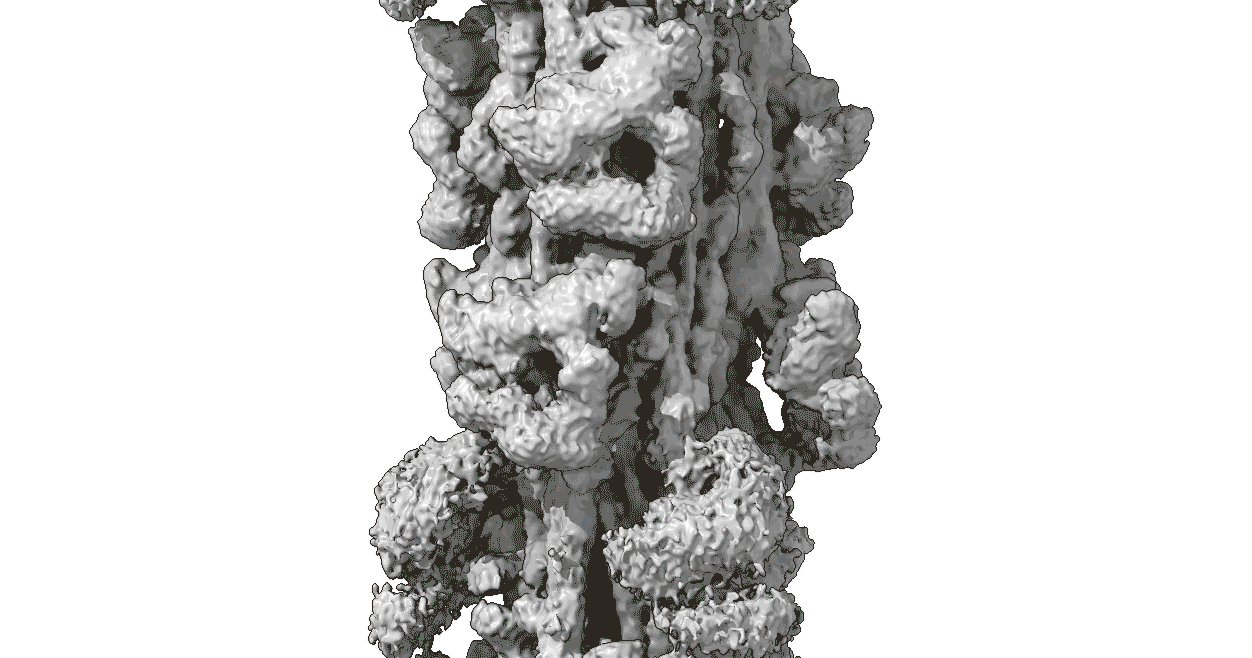
Movement is a defining feature of life. Our goal is to understand how muscles contract to move the body, and how cells move and change shape. We use cryo-electron microscopy and image analysis to study the structure of the motor proteins and filaments that underlie these movements. We are especially interested in the molecular switches that turn contraction and motility on and off. We study skeletal, cardiac, smooth and nonmuscle cells, and complement this work with low-angle X-ray diffraction of muscle and solution biophysical techniques. Our studies provide a structural basis for understanding diseases that result from mutations in contractile proteins.
Read moreApproach: cryo-EM
Advances in Cryo-EM Microscopy with Titan Krios
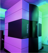
Understanding biological function requires knowledge of the three-dimensional structures of the macromolecular complexes that power the cell. Recent, revolutionary advances in cryo-electron microscopy make it possible to observe protein structure at near-atomic resolution, both in purified complexes and in the cell. We use cryo-EM (together with other EM and X-ray diffraction techniques) to determine the structures of the protein molecules of muscle, and the higher order complexes that they form, in different functional states. We make use of the superbly equipped cryo-EM Core at UMass Chan Medical School (Massachusetts Facility for High-resolution Cryo-electron Microscopy) to carry out this work.
Meet the LabPublications
Recent Publication History
Somavarapu AK, Ge J, Yengo CM, Craig R, Padron R. Cryo-EM Reveals How Cardiomyopathy Therapeutic Drugs Modulate the Myosin Motors of the Heart. bioRxiv. 2025:2025.10.29.685122. doi: 10.1101/2025.10.29.685122.
Nguyen V, Gautam R, Somavarapu AK, Dutta D, Patra A, Ge J, Yengo CM, Padrón R, Craig R. “QuickStainer”: a rapid negative staining device for improved preservation of molecular structure. bioRxiv. 2025:2025.10.11.681814. doi: 10.1101/2025.10.11.681814.
Dutta D, Kim Y, Ho C, Investigators S, Seidman JG, Seidman CE, Craig R, Padrón R. Thick filament molecular interfaces play a critical role in pathogenesis of hypertrophic and dilated cardiomyopathy. bioRxiv. 2025:2025.10.03.680256. doi: 10.1101/2025.10.03.680256.
Koubassova NA, Dutta D, Ma W, Tsaturyan AK, Irving T, Padrón R, Craig R. Annotating the x-ray diffraction pattern of vertebrate striated muscle. Biophys J. 2025;124(21):3663–77. Epub 20250915. doi: 10.1016/j.bpj.2025.09.019. PubMed PMID: 40954998; PMCID: PMC12694897.
Craig, R., Dutta, D., Koubassova, N. A., Tsaturyan, A. K. & Padron, R. (2025). Comparing cryo-EM structures of the vertebrate cardiac muscle thick filament. Biophys. Rev. In Press.
Publications